Urinary stones (urolithiasis), cause, treatment, prevention

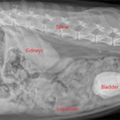
The development of stones in the urinary tract (either kidney or bladder stones) is referred to as urolithiasis. Like in humans, the kidneys of dogs are organs that filter blood through a thick network of tissue. Waste products are removed from the blood by the kidneys and are then transported via tiny, thin tubes called ureters in the belly. These waste components from the urine are sent by the ureters into the bladder, which serves as a holding tank for the waste to be collected as urine. The urethra, a bigger tube, is where the pee is afterwards emptied.
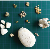
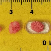
Urinary calculi, commonly referred to as uroliths or urinary stones, can develop in the bladder or kidneys. They could enter and obstruct the urethra, which is the larger tube that connects the bladder to the outside of the body, or the ureters, which are the paired tubes that connect the kidneys to the bladder. Uroliths can be made of a variety of materials. Strucvite (also known as "triple phosphate") and calcium oxalate uroliths are the two most prevalent forms of uroliths in dogs. Additionally, calcium phosphate, cystine, urate, or xanthine can be found in uroliths. Struvite uroliths are frequently associated with nutritional variables (high-ash, low-acid diet) and urinary tract infections in dogs.
Dogs, especially male, may experience urethral blockage as a result of "plugs" composed of small uroliths and other materials. specific forms of uroliths are more common in specific dog breeds. Certain disorders and hereditary metabolic abnormalities lead to an abnormally high concentration of a chemical in the urine, which can precipitate into the form of a urolith. As a result, the likelihood of urolith development is increased. For instance, pets with a birth abnormality known as portosystemic shunt may develop urate uroliths.
Signs
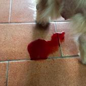
The symptoms of uroliths in the bladder or urethra include straining to urinate, often urinating little amounts of urine (pollakiuria), and having blood in the urine (hematuria). Nevertheless, since other conditions might cause similar symptoms, it is crucial to have your dog or cat examined by a veterinarian if any of these signs appear. Not all dogs with these symptoms have kidney stones. Sometimes, pets with uroliths may not exhibit any symptoms at all, and the uroliths are only discovered by coincidence (incidental detection) when testing (such x-rays) are being done for other issues.
Can be emergency
Urethral obstruction, or complete blocking of the bladder's outflow by a urolith, is a medical emergency. Uremia is a build-up of waste materials in the body that would typically be expelled in the urine. If urine cannot be expelled from the body for 24 hours or more, the pet may die. If the urine's exit not obstructed , uroliths are not as serious. Notably, straining to urinate is the primary symptom that distinguishes urolithiasis causing urethral obstruction from urolithiasis that does not obstruct urine outflow. Pets with urethral blockage typically strain to urinate, but as a result, no urine flows out because the path of urine flow is blocked.
This condition causes weakness, lethargy, and finally, over the course of 24 to 48 hours, a coma and death if ignored.
Causes
Urinary stones, also known as uroliths or bladder stones, can form in the urinary tract of dogs due to various factors. The exact cause can vary depending on the type of stone formed, but some common factors include:
- Diet: The type of diet a pet consumes can play a significant role. Certain minerals in the diet can lead to the formation of crystals, which can then aggregate and form stones. For example, struvite stones often form in animals with urinary tract infections that make the urine alkaline, promoting the formation of these crystals.
- Water Intake: Insufficient water intake can lead to concentrated urine, making it easier for crystals and stones to form. Pets that do not drink enough water are at a higher risk.
- Breed Predisposition: Some breeds are genetically predisposed to forming certain types of stones. For instance, Dalmatians are known to be prone to urate stones.
- Urinary Tract Infections (UTIs): Infections can create an environment conducive to crystal formation and stone aggregation. Bacterial presence can alter the pH of urine, encouraging the formation of certain types of stones.
- Urinary Tract Abnormalities: Structural issues in the urinary tract can cause urine to stagnate, allowing crystals and stones to form more easily.
- Metabolic Disorders: Certain metabolic conditions, like hypercalcemia (high levels of calcium in the blood) or hyperparathyroidism, can predispose animals to stone formation.
- Obesity: Overweight pets are more prone to urinary issues, including the formation of stones.
- Lack of Exercise: Physical inactivity can contribute to various health problems, including urinary issues.
- Medications: Some medications can lead to the formation of certain types of stones as a side effect.
- Genetic Factors: There might be genetic factors at play, making some animals more susceptible to stone formation than others.
Treatment
The treatment of urinary stones in dogs depends on the type of stone, its size, location, and the overall health of the animal. Here are some common approaches to treating urinary stones in pets:
- Dietary Management:
- Prescription Diets: Specialized prescription diets are available for dissolving certain types of stones. These diets work by altering the pH of the urine and reducing the concentration of stone-forming minerals. For example, diets formulated to dissolve struvite stones or prevent their formation are often prescribed.
- Hydration: Encourage your pet to drink more water. Increased water intake helps dilute the urine and may help prevent the formation of certain types of stones.
- Surgery: In some cases, especially when the stones are too large to pass naturally or are causing severe blockages or pain, surgical removal may be necessary. There are different surgical techniques, including traditional surgery and minimally invasive procedures like laser lithotripsy.
- Non-Invasive Techniques:
- Extracorporeal Shock Wave Lithotripsy (ESWL): This non-invasive technique uses shock waves to break stones into smaller pieces, making it easier for the pet to pass them naturally.
- Urethral and Cystoscopic Procedures: Certain stones can be removed through the urethra or cystoscope without major surgery.
- Medications:
- Pain Management: Pain medications may be prescribed to keep the pet comfortable, especially if the stones are causing pain or discomfort.
- Antibiotics: If there is a urinary tract infection associated with the stones, antibiotics may be prescribed.
- Lifestyle and Dietary Changes:
- Weight Management: If obesity is a contributing factor, weight loss through diet and exercise can be beneficial.
- Regular Exercise: Encouraging physical activity can improve overall health and may help prevent the recurrence of urinary issues.
- Monitoring and Follow-up: Regular veterinary check-ups and monitoring of the pet's urine are crucial to prevent the recurrence of urinary stones. This might include periodic urinalysis and imaging studies.
It's important to note that the appropriate treatment will be determined by a veterinarian after a thorough examination and diagnostic tests. If you suspect your pet has urinary stones or if you're concerned about their urinary health, consult a veterinarian promptly for proper diagnosis and tailored treatment options. Early intervention and appropriate management can significantly improve the outcome for pets with urinary stones.
Prevention
Prevention strategies often involve dietary management, ensuring pets have access to fresh water at all times, and addressing underlying health conditions. If you suspect that your pet may have urinary stones or if you're concerned about their urinary health, it's essential to consult a veterinarian for proper diagnosis and guidance on treatment and prevention.
Since there is no one symptom that is 100% specific to urolithiasis, testing is routinely done on patients who are suspected of having uroliths. The most often performed tests are routine blood work (complete blood count and serum biochemistry panel), urine sample analysis (urinalysis), and diagnostic imaging (x-rays and/or ultrasound).
Urine sample analysis may reveal the presence of blood or crystals (albeit the latter are not always related to the former).


